Dear Editor,
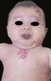
PHACE syndrome (OMIM: 606519) is a neurocutaneous syndrome whose cardinal sign is the presence of infantile hemangioma located on the face, neck, or scalp accompanied by at least one of the following extracutaneous manifestations included in its acronym: posterior fossa malformations, hemangiomas, arterial lesions, cardiac abnormalities/coarctation of the aorta, eye anomalies.1 Patients with PHACE syndrome commonly seek medical care for treatment of the facial angiomatous lesions and the respiratory symptoms arising from subglottic or mediastinal angiomatous lesions.2 However, recent case reports suggest a potential link between PHACE syndrome and hearing loss.3 A a four-year old female patient with a diagnosis of PHACE syndrome presented with ipsilateral hearing loss and decreased visual acuity. There was a malformation in the posterior fossa, but without cognitive deficits or other abnormalities. The patient received multidisciplinary follow-up. Physical examination revealed cranial asymmetry. The angiomas were characterized as segmental hemangioma of infancy interspersed with apparently normal skin. The lesions extended across the left hemiface, upper lip, left cervical region, left shoulder, and upper left thorax (Figure 1). Oroscopic examination showed an angiomatous lesion on the palate (Figure 2). Otoscopic examination on the left revealed a narrowed external auditory canal, and the tympanic membrane (TM) was intact but atrophic and with an angiomatous lesion (Figure 3). Auditory brainstem response (ABR) revealed a left-side conductive hearing loss of 50 dB, and tympanometry, for assessing the integrity of the middle ear and TM, found a type B curve. These findings confirmed this alteration in the middle ear as the cause of the hearing loss, suggesting otitis media with effusion, a common diagnosis in the pediatric population. We thus opted for a left myringotomy with placement of a TM ventilation tube to drain the secretion. During the procedure, effusion with a typical appearance was identified in the middle ear. However, even after the surgical procedure, and at subsequent follow-up visits, the patient maintained her left-side conductive hearing loss, as well a complaint of low school performance. We ordered a CT scan of the ears and mastoids, which is not a common routine test for the identification of conductive hearing loss in childhood, and it showed a lesion with the material density of soft tissues in the anterior and superior portion of the left middle ear, consistent with an angiomatous lesion. Other surgical options were ruled out. The patient was referred for fitting a hearing aid and continues in outpatient follow-up. The criteria for confirming definitive and possible PHACE syndrome, distributed in major and minor criteria, involve a long list of disorders, including arterial, cerebral, cardiovascular, ocular, midthoracic, and abdominal ano-malies.1 The diagnosis of definitive PHACE syndrome requires the presence of a hemangioma of infancy greater than 5cm in diameter on the head (including the scalp), plus one major or two minor criteria, or angioma on the neck, upper thorax, or thorax and upper limbs, plus two major criteria.1 The patient described here met the criteria for definitive PHACE syndrome based on the presence of an angiomatous lesion on the face and the posterior fossa anomaly (dysgenesis of the left cerebellar hemisphere). The patient’s ocular alterations (divergent strabismus and left myopia) are not part of the clinical findings related to the syndrome. The association of PHACE syndrome with hearing loss only appeared in the medical literature in 2010, when just a few cases of this association were published, including hearing losses involving both the middle ear and areas of the cochlea and cochlear nerve, that is, both conductive and neurosensory losses.3 The case described here was a conductive hearing loss with middle ear involvement due to two simultaneous factors: otitis media with effusion and an angiomatous lesion. It is known that infantile hemangiomas may either be present at birth as precursor lesions or only manifest some weeks later, and that hearing loss may manifest after this time. Therefore, normal hearing tests at birth do not guarantee normal hearing throughout childhood. Thus, in patients with a diagnosis of PHACE syndrome, we believe that the classically recommended care should also include monitoring of hearing acuity.


