Dear Editor,
Congenital smooth muscle hamartoma is defined as a rare asymptomatic benign skin defect detected in newborns and young children. Clinically it manifests as a hyperpigmented or skin-colored plaque where prominent vellus hairs may be observed.1,2 The condition is characterized by the proliferation of smooth muscle bundles in the dermis, which can reach the hypodermis where a connection to the hair follicle may occur.1,3 Due to its atypical manifestation, it may be confused with other cutaneous diseases, thus histological confirmation is necessary.1-5
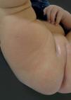
A 56-day-old Caucasian girl from São José do Rio Preto - SP, born with 33 weeks due to a cesarean twin birth, presented with a brownish macula in the posterior region of the right thigh that had appeared 19 days after birth. Physical examination revealed a hyperchromic plaque with discreet palpation, regular contours and absence of hair. Rubbing the lesion produced a transient erythema suggesting a pseudo-Darier's sign (Figure 1). Initially, the diagnostic hypotheses included: solitary mastocytoma, Becker's nevus, pilar leiomyoma, and smooth muscle hamartoma. We performed an ultrasound of the soft parts of the lesion that revealed a discrete obliteration of skin and subcutaneous cellular tissue, and absence of changes in the musculature. No images suggesting cystic or solid lesions were detected and no alterations of structures visualized by Doppler were observed. An incisional biopsy was performed, which revealed a proliferation of smooth muscle bundles that were randomly arranged in the reticular dermis. We did not observe atypia or inflammatory activity and the histological findings were compatible with congenital smooth muscle hamartoma (Figure 2). Additionally, immunohistochemical tests revealed that the smooth muscle was positive for actin (Figure 3).
According to the literature, congenital smooth muscle hamartoma is a rare malformation with a higher prevalence in males that mainly affects the lumbosacral region, although it may also occur in the trunk, arms, thighs and buttocks.2,3,4 In view of this fact, this case is even rarer, since the patient was female and the lesion appeared on her thigh.
The diagnosis of congenital smooth muscle hamartoma is complex due to the clinical similarity with other diseases, such as Becker's nevus, pilar leyomioma, solitary mastocytoma, among others.1,2,3 The histological and immunohistochemical examinations were necessary for a definite diagnosis. Becker's nevus is a benign tumor acquired by the smooth muscle that also presents muscle bundle hyperplasia, but an increase in the number of melanocytes, an enlargement of papillary cones and acanthosis is observed throughout the epidermis.3 Due to the histological similarity with smooth muscle hamartoma, a thorough medical history is necessary to distinguish the two conditions: Becker's nevus is not congenital, presents hypertrichosis and is typically located on shoulders and thorax.
Another differential diagnosis is the pilar leiomyoma, which presents a grouped proliferation of muscle bundles between the collagen fibers. The incisional biopsy preformed in our patient revealed smooth muscle bundles randomly arranged in the dermis, which is characteristic of smooth muscle hamartoma.
A diagnostic clue in our case was the pseudo-Darier's sign, which is present in 50% of cases of smooth muscle hamartoma.2,3 It appears after rubbing the lesion, due to the contraction of the piloerector muscle, and is characterized by skin indentation, piloerection, and color accentuation in a fleeting way.2 The true Darier's sign, which occurs in mastocytoma, is different as it is characterized by the onset of an erythema that arises after an injury stimulus and persists for one to two minutes due to the longer duration of histamine release and vasodilatation. 1
The diagnosis of muscle hamartoma should be considered before any congenital hair lesion and should be confirmed by biopsy. The absence of hypertrichosis, as occurred in our case, makes the diagnosis even more difficult as it can be confused with the pathologies mentioned above.
Therapeutic intervention is not necessary since congenital smooth muscle hamartoma is a benign tumor of the skin that is not associated with systemic manifestations and is not at risk of becoming malignant.1-3 The patient will be kept in clinical follow-up. □
AcknowledgementsWe would like to thank the pathologist Jorge Alberto Thome.


