Journal Information
Vol. 95. Issue 1.
Pages 112-113 (1 January 2020)
Visits
6175
Vol. 95. Issue 1.
Pages 112-113 (1 January 2020)
Case Letter
Open Access
Papillary syringocystadenoma in an uncommon location
Visits
6175
Giovanna de Araujo Horcel
Corresponding author
giohorcel@hotmail.com

Corresponding author.
, Juliana Milhomem, Samuel Henrique Mandelbaum, Rodrigo Ieiri
Dermatology Service, Santa Casa de São José dos Campos, São José dos Campos, São Paulo, SP, Brazil
This item has received

Under a Creative Commons license
Article information
Full Text
Bibliography
Download PDF
Statistics
Figures (3)
Show moreShow less
Full Text
Dear Editor,

Papillary syringocystadenoma is a rare neoplasm of sweat glands, which is present at birth in 50% of cases.1 It has predominantly apocrine differentiation, although an eccrine origin has been described in some reports.2,3

It usually presents as papule or plaque with a crustal surface that occurs almost exclusively in the head and cervical region.4

This report details the clinical observations as well as the dermatoscopic and histopathological findings of a case in an unusual location.

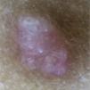
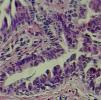
A 6-year-old male, a native and resident of São José dos Campos, SP, presented a lesion with progressive growth five years ago on the left flank. At the dermatological examination, a papule of pink-erythematous coloration was observed, with a smooth surface and a fibroelastic consistency, measuring 5mm×3mm (Fig. 1). Dermoscopic examination showed rounded structures of whitish-yellow color separated by whitish linear structures on an erythematous background (Fig. 2). The patient was referred for excision. Histopathological examination revealed the following: cystic invaginations covered by cells, sometimes squamous and sometimes columnar, with papilliferous projections to the light; tubular glands with large lights, covered by apocrine cells (Fig. 3).

Figure 1.

Rose-colored papule on the left flank.

(0.03MB).
Figure 2.

Dermoscopic examination showing rounded yellowish-whitish structures separated by linear whitish structures on an erythematous background.

(0.04MB).
Figure 3.

Histopathological examination: cystic cavity coated by basal cuboidal cells and columnar apocrine cells (Hematoxylin & eosin, ×400).

(0.12MB).

Papillary syringocystadenoma (SCAP) is a rare adnexal tumor most often derived from apocrine cells.3,4 It predominates in children and adolescents, and is observed at birth in 50% of cases,1 which differs from the case reported above.

In 75% of cases, it is located on the head or cervical region. Some cases in other topographies have already been described (scrotal region, vulva, back, abdomen, and axilla). These locations, as well as that of the case described, are even rarer. When located on the scalp, it may be associated with the sebaceous nevus of Jadassohn.4

Despite variable clinical presentation, a papule plaque is the most commonly found lesion type. In the majority, it is asymptomatic, but it can present pruritus, pain, and/or bleeding; it usually presents progressive growth, as in the case described.1,4

To date, articles on the dermoscopic findings of SCAP are scarce. There is a description of a horseshoe vascular pattern, which was not observed in the present case.3,5

SCAP can infect, bleed, ulcerate, and, in rare cases, it can progress to basal cell carcinoma (9%) or papillary syringo-cystadenocarcinoma. For these reasons, it was decided to perform the excision.5

Financial support

None declared.

Authors’ contribution

Giovanna de Araujo Horcel: Conception and planning of the study; composition of the manuscript.

Juliana Milhomem: Conception and planning of the study.

Samuel Henrique Mandelbaum: Critical review of the literature; critical review of the manuscript.

Rodrigo Ieiri: Participation in the design of the study.

Conflicts of interest

None declared.

References
[1]
E. Karg, I. Korom, E. Varga, G. Ban, S. Turi.
Congenital syringocystadenoma papilliferum.
Pediatr Dermatol, 25 (2008), pp. 132-133
[2]
T.C. Townsend, A.R. Bowen, K.K. Nobuhara.
Syringocystadenoma papilliferum: an unusual cutaneous lesion in a pediatric patient.
J Pediatr, 145 (2004), pp. 131-133
[3]
C.B. Bruno, F.N. Cordeiro, F.E.S. Soares, G.H.S. Takano, L.S.T. Mendes.
Aspectos dermatoscópicos do siringocistoadenoma papilífero associado a nevo sebáceo.
An Bras Dermatol, 86 (2011), pp. 1213-1216
[4]
A.P. Jalkh, A.C. Menezes, A. Gadelha.
Siringocistoadenoma papilífero localizado na coxa.
Surg Cosmet Dermatol, 5 (2013), pp. 273-275
[5]
Y.S. Einecke, E.B. Pinto, S.O. Silveira, M.A. Santos, A.M. Mendes, F.R. Carneiro.
Siringocistoadenoma papílifero congênito.
Rev SPDV, 76 (2018), pp. 79-82

How to cite this article: Horcel GA, Milhomem J, Mandelbaum SH, Ieiri R. Papillary syringocystadenoma in an uncommon location. An Bras Dermatol. 2020;95:112–3.

Study conducted at the Dermatology Service, Santa Casa de São José dos Campos, São José dos Campos São Paulo, Brazil.

Download PDF
Idiomas
Anais Brasileiros de Dermatologia
Article options
Tools
en pt
Cookies policy Política de cookies
To improve our services and products, we use "cookies" (own or third parties authorized) to show advertising related to client preferences through the analyses of navigation customer behavior. Continuing navigation will be considered as acceptance of this use. You can change the settings or obtain more information by clicking here. Utilizamos cookies próprios e de terceiros para melhorar nossos serviços e mostrar publicidade relacionada às suas preferências, analisando seus hábitos de navegação. Se continuar a navegar, consideramos que aceita o seu uso. Você pode alterar a configuração ou obter mais informações aqui.